UPWr Scientist studies and promotes a non-invasive method of animal diagnostics
Is a careful examination of a horse using a thermal imaging camera enough to diagnose diseases or injuries? Yes, and prof. Maria Soroko-Dubrovina from UPWr, who conducts research on this subject, has no doubts that this is the future in veterinary medicine and horse rehabilitation.
Thermography is a diagnostic method which uses the non-invasive registration of infrared radiation energy emitted from the surface of the body and transforming it into a temperature value. – Any increase in temperature means inflammation and with thermography we can detect it, even in the subclinical state of inflammation, when the animal doesn't show visible symptoms. A thermal imaging camera is a sensitive device that can detect temperature changes up to 0.1º C. Thanks to this, we are able to show the pathological changes occurring in the animal's body before they fully develop – says Prof. Maria Soroko-Dubrovina from the Institute of Animal Breeding, UPWr, who has been working with thermography for fourteen years.
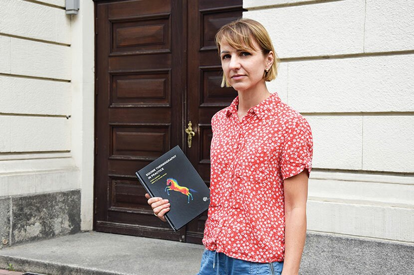
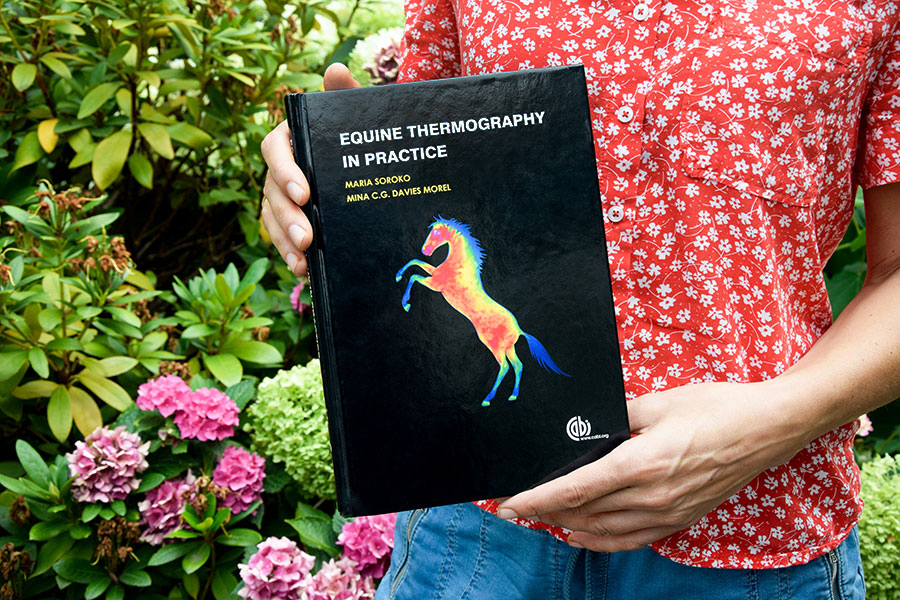
Her book "Equine Thermography in Practice'' is the result of her interests. It was published by the British publishing house CABI. The book was a great success, it was sold in 30 countries around the world in over 600 printed copies and 75 ebooks. It has just been reprinted on the shelves of the bookstore, and a second edition is planned for this year.
Random interest, conscious research
Prof. Soroko-Dobrovina's interest in thermography began during her second-cycle studies at Aberystwyth University by chance. Access to thermographic equipment made her decide to conduct research on the use of thermography to diagnose the cause of lameness in racing horses.
– I developed these interests during my doctoral studies at the Faculty of Biology and Animal Science, at the Wrocław University of Environmental and Life Sciences. The research focused mainly on the use of thermography in assessing the impact of training on changes in body surface temperature in racing horses. Since completing my PhD, I’ve been carrying out further research using thermography in equine physiotherapy (in assessing the effects of physical treatments), in assessing the level of stress, in equestrianism (e.g., in fitting equestrian equipment). I’m also expanding my research on the use of thermography in other animals, such as cattle, poultry, dogs – she says.
The book "Equine Thermography in Practice" by prof. Soroko-Dubrovina has been sold in 30 countries around the world photo by Martyna Kostrzycka
The UPWr scientist admits that the use of thermography for diagnosing horses in scientific research is nothing new. Its first use in equine veterinary medicine took place in the mid-1960s by American scientists. Based on the knowledge gained in medical thermography, they demonstrated the usefulness of this technique in detecting inflammation of orthopedic diseases, which were also confirmed by a radiological diagnosis. A decade later, its usefulness in detecting early inflammation up to 14 days before clinical inflammation was revealed for the first time.
– However, the limitations of this technique at that time, such as the low sensitivity of the equipment and the lack of qualified experts in the field were the source of many diagnostic errors, contributing to the abandonment of this technique in veterinary practice. Only at the turn of the 1980s and 1990s there was a significant technological advance in the apparatus and its renewed interest in the research world of horses – explains Prof. Soroko-Dubrovina, adding that this method has many advantages.
It helps to locate the site of the injury, which facilitates further diagnosis by a veterinary doctor. It is also a useful tool in the practice of a trainer or physiotherapist. It helps to assess the effects of physiotherapeutic procedures by locating areas with altered blood supply due to muscle and tendon soreness or overloading and the accompanying tension. For the practice of farriers, thermography is used to determine the work of both forged and bare hooves, or to locate the inflamed area of the hoof. – And above all, it is non-invasive, so it can be performed safely on any horse, regardless of its health condition – highlights the professor.
This site uses custom cookies to ensure that it functions properly. Some are necessary for the page to run, so will always remain active. These cookies will store information about the user's cookie settings. In addition, third-party cookies are used for external tools. For more information see the privacy policy.
Purpose
Enables storage (such as cookies) related to advertising.
Agreed
Sets consent for sending user data to Google for online advertising purposes.
Agreed
Sets consent for personalized advertising.
Agreed
Enables storage, such as cookies (web) or device identifiers (apps), related to analytics, for example, visit duration.
Agreed
Enables storage that supports the functionality of the website or app, for example, language settings
Agreed
Enables storage related to personalization, for example, video recommendations
Agreed
Enables storage related to security such as authentication functionality, fraud prevention, and other user protection